3. Dioxin Research
3.1.1.
As Dr. Nancy Kerkvliet of OSU recently said,
    DECADES OF DIOXIN RESEARCH GENERATE MORE QUESTIONS THAN ANSWERS. Really, after years of study ("dioxins" give
    6,000,000 results in Google) and millions of dollars spent on research, scientists are not much closer today than they were 40+
    years ago to understanding the human health risks of exposure to dioxin. One problem in unraveling the mystery of dioxin has
    been the difference in species' response to dioxin exposure in animal studies. Extremely inconsistent findings in animal studies
    make it almost impossible to extrapolate animal study findings to humans, Kerkvliet said. There also is controversy surrounding
    how dioxin molecules affect the cells of animals and humans".
2,3,7,8-Tetrachlorodibenzo-p-dioxin (TCDD) is a toxic byproduct of various industrial, combustion, and natural processes that is now a persistent and ubiquitous environmental contaminant in air, water, soil, and food. TCDD exerts a broad spectrum of toxic effects even at very low concentrations. Lethal doses of TCDD cause slow death as a result of a wasting syndrome, while reproductive and developmental effects, tumor promotion, immune suppression, and skin lesions called chloracne are observed at nonlethal doses. TCDD is probably best known as a contaminant in Agent Orange, a mixture of N-butyl esters of 2,4-dichlorophenoxyacetic (2,4-D) and 2,4,5 trichlorophenoxyacetic (2,4,5-T) acids widely used as defoliants by the United States during the war in Vietnam (1962–1971). In 1968, a mass food poisoning termed “Yusho” occurred in western Japan, and the causal agent of “Yusho” was thought to be polychlorinated dibenzofurans (PCDFs) [65]. In 1976, a severe industrial accident occurred in Seveso, Italy, and up to kilogram quantities of TCDD and additional TCDD-like chemicals were dispersed into the air, with adverse health consequences for the local population [30]. In 2004, Viktor Yushchenko, who was the president of Ukraine at that time and had been exposed to TCDD at a single oral dose 5 million-fold higher than the accepted daily exposure for the general population, became a widely recognized victim of TCDD poisoning. Those rare cases of high levels of dioxin exposure are presented in this Figure.
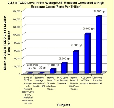
Wide variety of health effects have been traditionally linked to high exposure to dioxins, so the common is that risk assessments based on extrapolating high-dose effects such as cancer may have over-estimated the toxicity of dioxins at low levels. Their more widespread application in toxicology dates back only 15 years or so to models In physiologically based pharmacokinetic (PBPK) models developed for polychlorinated biphenyls and other persistent lipophilic compounds.
However, with a lot of uncertainty about dioxin's effects on humans and animals, that hasn't stopped the Environmental Protection Agency from issuing strict guidelines for allowable human exposure to the chemical. The first in-depth EPA study of dioxin, released in 1985, established acceptable exposure levels of dioxin for humans. These levels were considered excessively low by some scientists and at the urging of several large U.S. corporations, the EPA conducted a lengthy review of its first study of dioxin. The second EPA report on dioxin was far from calming worries about the chemical, the report heightened concerns by announcing that the safety margin for exposure to dioxin was much smaller than scientists first thought. The EPA review added that dioxin effects on the immune system, the reproductive system and disruption of hormones might even be greater than the risk of cancer from exposure to dioxin. In practical terms, that finding meant that EPA scientists were no longer talking about levels of safe exposure to dioxin. They were saying that exposure to the chemical at any level is potentially harmful to human health and to the environment in general.
Many scientists representing US chemical industry disagree with several of the assumptions the EPA made in arriving at this conclusion, as earlier scientists hired by Philip Morris rejected any carcinogenic effects from cigarette smoking. The EPA's current position dictates how the chemical will be regulated, and industries that generate dioxin as a by-product - such as paper and pulp mills, trash incinerators as well as incinerator for medical and industrial wastes. Here - our meeting with Prof. A. Schecter (in the photo) coming to USSR to collect samples of mother milk from dioxin-contaminated areas in Siberia.

3.1.2. Studies on the Rodents Treated with PAHs vs TCDD
Since the mid-1980s, Tsyrlov's lab switched research priorities to molecular mechanisms of TCDD action and related medico-biological outcomes. Initially, in vitro and in vivo human-relevant model systems were utilized to evaluate the inter- and intra-species variation in catalytic activities of CYP1A1 and CYP1A2 induced with polycyclic aromatic hydrocarbons (PAHs) and TCDD. Thus, in liver microsomes of TCDD-treated rats, Tsyrlov with Olga Chasovnikova Olga Chasovnikova.pdf revealed rapidly synthesized Cyp1a1 with higher aryl hydrocarbon hydroxylase (AHH) molecular activity than that of Cyp1a1 in 3-MC-treated rats.[23] In another study, Tsyrlov with Tatiana Duzchak With Duzhak.pdf used mutually-depleted antibodies and monoclonal antibodies developed against murine and rat Cyp1a1 and Cyp1a2. Namely, enzyme kinetics, spectral and electrophoretic parameters, immunoblotting, Ouchterlony double immunodiffusion and immunoinhibition analyses of purified CYPs were compared to CYPs in microsomes from benzo[a]pyrene (B[a]P)-treated Wistar rats and C57BL/6 mice.[24] Conclusions drawn from these and other studies allowed to incline toward mice rather than rats as an animal model for the CYP1A1/CYP1A2 in humans. Also, B[a]P and 2,3,7,8-TCDD were shown as xenobiotics that cause persistent adverse health effects in humans. Tsyrlov's team conducted studies on long-term effects of B[a]P on AHH activity during tumor appearance in mice homozygous and heterozygous for the Ahb allele and Ahd allele at the Ah gene locus,[25] a B[a]P protracted induction of AHH and mutagen effects in lymphocytes from workers of the aluminum industry, where level of exposure to B[a]P is up to 100 μg/m,[26]. Also, with Vladimir Ostashevsky V Ostashevsky.pdf and Konstantin Gerasimov, Tsyrlov measured the AHH and mitogenic index in lymphocytes, placenta, and liver biopsies from South Vietnamese previously exposed to 2,3,7,8-TCDD and its congeners, - the components of an Agent Orange.[27] Also, in the 1980s Tsyrlov has prepared two monographs (one with Vyacheslav Lyakhovich), both on mammalian CYPs induced by barbiturates, PAHs, polychlorinated biphenyls or dioxins and dioxin-like compounds (DLC), and applied medico-biological implications.[28],[29].
3.1.3.
An especial paper was published in 1986 in the major academic national journal,[30] which still keeps itself to itself. B[a]P was ingested by mice with allelic differences at the Ah locus, which reproduced the "first pass metabolism" model of Daniel Nebert (summarizing in[31]). To epitomize the model from the point of view of pluripotent hematopoietic stem cells (PHSC), Tsyrlov collaborated with the established stem cell researcher, Dr. Irena Tsyrlova (in the photo, Drs. Tsyrlov and Tsyrlova with Dr. Paul Thomas of Rutgers University).
 A primary immune response, measured by the number of IgM antibody-forming cell (AFC), revealed a drastic difference between C57BL/6 (genotype Ahb/Abb) and DBA/2 (genotype Ahd/Abd) mice, with adjustment for bone marrow aplasia characteristic for B[a]P-ingested DBA/2 mice. As aplasia was time-reversible, the model was considered an immunologically unique alternative to the classic model[32] where murine PHSCs are detected by injecting bone marrow cells into irradiated recipients. Moreover, it was registered for the first time that more than 70% of PHSCs from B[a]P-ingested mice with Ahd/Abd genotype are involved in proliferation and that a disproportional shift of PHSC into S phase of the cell cycle is accompanied by stimulation of erythropoiesis and granulocytopoiesis to the detriment of an immunogenesis. The very same feature has been described in the mid-2000s for clonal hematopoiesis of indeterminate potential (CHIP) in older people especially those who had been exposed to environmental mutagenic pollutants like B[a]P. The CHIP is associated with an increased risk of hematologic cancer and atherosclerotic cardiovascular disease.[33] So, the data obtained in 1986 have made the pioneer contribution to a current major medical field, as the CHIP is considered the most important discovery in cardiology since statins.
REFERENCES
        [23] Tsyrlov, I.B., Chasovnikova, O.B., Grishanova, A.Yu., Lyakhovich, V.V. (March 1986). “Reappraisal of the liver benzpyrene hydroxylase synthesized de novo after treatment of rats with 2,3,7,8-tetrachlorodibenzo-p-dioxin and 3-methylcholanthrene”. FEBS Letters 198:225-228. DOI:10.1016/0014-5793(86)80410-0
        [24] Tsyrlov, I.B., Duzchak, T.G. (November 1990). “Interspecies features of hepatic cytochromes P450 IA1 and P450 IA2 in rodents”. Xenobiotica 20(11):1163-1170. DOI:10.3109/00498259009046836
        [25] Tsyrlov, I.B., Lyakhovich, V.V. (April 1979). “A comparison of the activities of aryl hydrocarbon monooxygenase in liver microsomes from mice of different strains during prolong 3,4-benzo(a)pyrene administration”. Biochimica et Biophysica Acta 584:11-20. ISSN: 0304-4165
        [26] Kessova, I.G., Sedova, K., Tsyrlov, I.B. (December 1990). Evaluation of monooxygenases catalyzing activation of carcinogens, and evaluation of mutagenic effects of benzo[alpha]pyrene in blood lymphocytes from workers of aluminum industry”. Oncology 13:12-15. ISSN:0378-584X
        [27] Tsyrlov, I.B., Ostashevsky, V., Gerasimov, K.E., Rumak, V.S. (July 1990). “Characteristics of hepatic and lymphocyte monooxygenases in South Vietnam’ people with chlorophenoxy herbicides exposure”. Organohalogen Compounds 1:305-308. ISSN:1026-4892
        [28] Lyakhovich, V.V., Tsyrlov, I.B. (February 1981). “Индукция Ферментов Метаболизма Ксенобиотиков” (in Russian). “Induction of Xenobiotic Metabolizing Enzymes”. 239 pp. Ed. Academician A. Yasaitis. Novosibirsk, Nauka Publishing House. ISBN: MH-05082
        [29] Tsyrlov, I.B. (December 1990). «Хлорированные Диоксины: Биологические и Медицинские Аспекты» (in Russian). “Polychlorinated Dioxins: Biological and Medical Aspects”. 216 pp., Ed. Academician V. Vlassov. Novosibirsk, Nauka Publishing House. ISBN: MH-10270
        [30] Tsyrlova, I.G., Kozlov, V.A., Tsyrlov, I.B. (April 1986). “A new experimental model for studying bone marrow cells participation in antibody genesis”. Cytology 28:102-106. UDC:57.083.3:612.419
        [31] Nebert, D.W., Shi, Z., Galvez-Peralta, M., Uno, S., Dragin, N. (September 2013). “Oral benzo[a]pyrene: understanding pharmacokinetics, detoxication, and consequences – Cyp1 knockout mouse lines as a paradigm”. Molecular Pharmacology 84:304-313. DOI:10.1124/mol.113.086637
        [32] Till, J.E., McCulloch, E.A. (February 1961). “A direct measurement of the radiation sensitivity of normal mouse bone marrow cells”. Radiation Research 14:213-222. DOI:10.2307/3570892
        [33] Jaiswal, S., Natarajan, P., Silver, A.J., Gibson, C.J., … Ebert, B.L. (total of 22 authors) (July 2017). “Clonal hematopoiesis and risk of atherosclerotic cardiovascular disease”. The New England Journal of Medicine 377:111-121. DOI:10.1056/NEJMoa170119
3.2. Mechanism-based models for induced expression of target genes by TCDD via AhR transcription complex
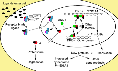
2,3,7,8-TCDD is known to trigger the cellular receptor AhR mediated (bHLH)/Per-Arnt-Sim(PAS) domain superfamily of proteins, which transcriptionally regulates the ''CYP1'' genes and involve in an array of genetic networks and subcellular processes critical to life.[34] Each of these processes is dependent on the binding of the AhR to the dioxin-response element, DRE (also known as XRE, the xenobiotic response element), the consensus core sequence of which is 5’-TNGCGTG-3’, within regulatory regions of the target gene. The cytoplasmic AhR exists in the absence of a ligand as a tetrameric complex composed of a 95-105 kDa ligand-binding subunit, a dimer of hsp90, and the immunophilin-like X-associated protein 2 (XAP2), also known as AH receptor-interacting protein, AIP. Despite the importance of the AhR in target tissues, the own transcriptional regulation of AhR and its cytosolic complex key members still remain to be established. To this point, a computational search for DRE sequences was undertaken among mammalians genes (including the AhR gene) - all calculated using a method of reverse position weight matrix constructed from bona fide DREs.[35] Therefore, the revelation of DREs in the genes encoding other members of bHLH/Per-Arnt-Sim(PAS) superfamily of proteins would shed a light on the potential mechanisms of expression, the stoichiometry of unliganded AhR core complex, and its degradation vs biosynthesis dynamics. Therefore Tsyrlov collaborated with bioinformatics experts at Laboratory of Theoretical Genetics, Institute of Cytology and Genetics, Russian Academy of Sciences to utilize the SITECON[36] for quantitative evaluation of DREs in genes encoding members of the AhR cytoplasmic complex. In contrast to having lower than 0.95 threshold position weight matrix-based methods, the SITECON proved to be a highly efficient ''in silico'' tool designed for recognition of potentially active transcription factor binding sites, TFBS, and is based on conservative conformational and physicochemical properties detected for a set of experimentally proven TFBSs, and was verified to assess functionally active binding sites in several mammalian genes, including promoter DREs in human CYP1A1 and CYP1B1 genes. In fact, three DREs were confirmed within downstream the TSS of the AhR gene, and at a 0.95 threshold, SITECON also revealed one additional upstream DRE (-7398 to -7385), which, despite its distant location from TSS, could be potentially active.[37] That proposed still not investigated the mechanism of AhR recovery from deep depletion due to 2,3,7,8-TCDD-activated AhR transcriptional signaling, and an over-expression of AhR in various malignant cells.[38] Even less was known about what factors regulate expression of genes that encode HSP90s, and no data existed as regard DREs within regulatory regions of XAP2 and ''ARNT'' genes, despite the fact that Tsyrlov and Pokrovsky had mentioned the latter in their paper in Xenobiotica in 1993 (https://www.wikigenes.org/e/gene/e/57491.html). To the point, Tsyrlov with Dmitry Oshchepkov (in the photo) and his colleagues bioinformaticians from Institute of Cytology and Genetics/RAS [37] detected five unknown active DREs within the regulatory region of the human XAP2 gene as well as the homologous gene in chimp, and every of DRE found was recognized with very high RS.

The above finding directly applied to the assumption of Yoshiaki Fujii-Kuriyama, namely that the level of susceptibility of the target gene to its inducer depends on a number of DRE motifs within the gene regulatory region.[39] Working with the mammalian gene models containing DREs in upstream activating sequence regions, Fujii-Kuriyama with colleagues found that models with tandem repeats of DREs in the promoter sequence of the CYP1A1 gene synergistically increased the transcriptional activity after the treatment by 3-MC, at median ED50 of 0.2 nM. So, as the XAP2 gene contains five promoter DREs, it might be significantly trans-activated by 2,3,7,8-TCDD at sub-nanomolar concentrations, which makes this chaperone protein a real regulatory factor for unliganded AhR, as XAP2 retains the AhR in the cytoplasm, protects AhR against labilization, ubiquitin degradation, and XAP2 over-expression might enhance cytosolic AhR content. Another essential molecular chaperone proteins, a dimer of HSP90s, interacts with the bHLH and PAS domains of AhR, which maintains the AhR in a conformation required for XAP2-AhR binding. To this point, SITECON mapped eight DREs in the regulatory region of the HSP90AA1 gene (of those six – upstream of TSS), all characterized by RS ranging from 0.959 to 0.988. Lastly, five DRE binding sites in the human ''ARNT'' gene were detected by the SITECON, each of which with a high RS number. The data on multiple DREs found in regulatory regions of genes encoding proteins, members of the AhR/XAP2/HSP90AA1 cytoplasmic complex, would elucidate their degradation vs biosynthesis dynamics and stoichiometry of unliganded AhR core complex. The most of newly and previously predicted DREs were found to be conservative in mammals. The human cell model was suggested by Tsyrlov and colleagues on an autoregulatory feedback loop maintaining constitutive and induced levels of the AhR in the cytoplasm.
REFERENCES
[34] Beischlag, T.V., Morales, J.L., Perdew, G.H. (May 2008). “The Aryl Hydrocarbon Receptor Complex and the Control of Gene Expression”. Critical Reviews in Eukaryotic Gene Expression 28:207-250. ISSN: 10454403
[35] Sun, Y.V., Boverhof, D.R., Burgoon, L.D., Fielden, M.R., Zacharewski, T.R. (August 2004). Comparative analysis of dioxin response elements in human, mouse, and rat genomic sequences. Nucleic Acids Research 32:4512-4523. DOI:10.1093/nar/gkh782
[36] Oshchepkov, D.Yu., Vitiaev, E.E., Grigorovich, D.A., Ignatieva, E.V., Khlebanova, T.M. (July 2004). “SITECON; a tool for detecting conservative conformational and physicochemical properties in transcription factor binding site alignments and for site recognition”. Nucleic Acids Research 32:208-212. DOI:10.1093/nar/gkh474
[37] Nedosekina, E.A., Oshchepkov, D.Yu., Mordvinov, V.A., Katokhin, A.V., Kuznetsova, T.N., Shamanina, M., Tsyrlov, I.B. (August 2007). “Detection of new potentially active DRE sites in the regulatory region of human genes encoding components of Ah receptor cytosolic complex”. Organohalogen Compounds 69(2):1890-1894. ISSN:1026-4892
[38] Murray, I.A., Patterson, A.D., Perdew, G.H. (December 2014). “Aryl hydrocarbon receptor ligands in cancer: friends and foe. Nature Reviews (Cancer) 14:801-814. DOI:10.1038/nrc3846
[39] Fujii-Kuriyama, Y., Sogawa, K., Mizukami, Y., Suwa, Y., Muramatsu, M., Kawajiri, K., Gotoh, O., and Tagashira, Y. (May 1985). “Molecular multiplicity and gene structure of microsomal cytochrome P-450 in rat liver”. Gann Monograph on Cancer Research 30, 157-169. ISBN-10: 03064212323.3. Studies on dioxins in Vietnam.
[Dioxins in Vietnam .pdf] (Dioxins%20in%20Vietnam%20.pdf)
Dioxins in Vietnam Book .pdf
Vietnamese and foreign scientists gathered at a conference in March 2006 to compare and verify scientific evidence of the debilitating effects of Agent Orange on victims. The conference in Hanoi represents an effort on behalf of all parties in attendance to fight the battle for rightful compensation for victims to the very end. In attendance were some 200 local and foreign scientists, activists and Vietnamese victims, double the number originally expected, reported Tuoi Tre newspaper.
    Experts speak up. According to Dr. Le Ke Son of Vietnam’s National Steering Committee for Agent Orange Impact Relief, the latest
    studies show US troops may have sprayed a whopping 86 million liters AO during the Vietnam War compared to the previous
    estimation of 72 million liters. Scientists have also detected gene and chromosome mutations to those exposed to AO/Dioxin,
    resulting in serious birth defects among offspring for generations, Son said. “We still need more research to convince people of the
    link between such deformities and AO/Dioxin, but the much higher incidence of children with congenital anomalies in AO affected
    areas is a testament to the deadly connection,” Tuoi Tre quoted Nguyen Van Tuong of the Hanoi Medical University as saying.
    Several Vietnamese did have normal kids but their children conceived after the war were acutely affected by the defoliant while
    many others gave birth to six or seven affected children in a row. “AO/Dioxin is the culprit,” he affirmed.

    Dr. Ilya B. Tsyrlov, who leads a study team from the US company Xenotox, Inc. added Agent Orange contains TCDD, one of the
    most stable and potentially carcinogenic compounds know to man. However, the list of dioxin-related diseases released by the US
    Science Academy has yet to include defects among the offspring of veterans,” Prof. Bernard Doray from France said, adding
    “studies on the toxicity have been distorted.” Although the US Government has taken some steps to relieve the impact of AO on
    the environment in Vietnam, it has denied responsibilities and has yet to offer compensation to AO-affected people in the country".
3.4. REGULATION OF HUMAN GENES OF INFLUENZA A VIRUS-ASSOCIATED NS1-BINDING PROTEIN BY TCDD.
Dioxin-linked mechanisms of avian H5N1 virus outbreak in South East Asia
3.4.1.
Agonist-induced recognition of a cognate DNA enhancer dioxin responsive element (DRE) does epitomize wide range of mammalian genes expression mediated via the Ah receptor pathway. The same was postulated also for viral DRE-containing genes expression caused by TCDD in infected human cells. In this study, such mechanistic concept applied to type A influenza virus nonstructural protein 1 binding protein (NS1BP) induction in humans and chicken. The data are presented at genetic, cellular, and population levels. Primers for mutation analysis were constructed for two DRE identified within enhancer region of the IVNS1ABP gene. Treatment of HeLa cell line with 0.1 nM of TCDD resulted in substantial increase of NS1BP protein level. This might add to influenza virus A non-structural protein 1 (NS1) inhibitory effect on cellular interferons, which determines antiviral resistance of emerging H5N1 virus. Recent H5N1 outbreaks among poultry in China and Vietnam might partially relate to chicken NS1BP, as outbreaks occurred in areas highly contaminated by dioxin-like compounds. Minimal dose of TCDD up-regulating human IVNS1ABP gene was estimated moderately above current TCDD blood level in general population. So, in human subgroups exposed to TCDD, its body burden might facilitate spreading of H5N1 if avian flu pandemic were to occur.
3.4.2.
Mechanistic studies on 2,3,7,8-TCDD (TCDD) numerous toxic effects in mammals have revealed mediatory role of the Ah receptor (AhR). TCDD-activated AhR was proven to up-regulate the expression of sensitive genes through direct gene transcriptional activation. That resulted from the nuclear AhR-Arnt heterodimeric complex binding to DRE in the 5’ flanking region of target gene.(1,2) Earlier we discovered that TCDD might also activate HIV-1 gene expression in infected human cells,(3) and later the AhR-mediated transcriptional pathway was demonstrated for activation by TCDD of CMV gene.(4) Mechanistic comprehension of gene activation by TCDD eventuated from DRE (core nucleotide sequence 3’ ACGCAC5’) identified in regulatory region of mammalian and viral genes.(1,2,5,6) In this study, the above mechanistic concept was applied to the IVNS1ABP gene encoding influenza virus A NS1 binding protein (NS1BP) in humans and chicken. Alongside an important role this protein plays in the virus life cycle, two DRE sequences were computationally detected in the IVNS1ABP 5’-flanking region, at positions - 7942 and - 687.(7)
The NS1 protein of influenza type A viruses (IAV) is known to prevent transcriptional induction of antiviral interferons (IFNs).(8) This protein also modulates host cell gene expression and inhibits double-strandedRNA (dsRNA)-mediated antiviral responses. The data suggest that the inhibition of splicing by the NS1 may be mediated by binding to NS1BP of the target cells.9 In this study, we treated human HeLa cells with TCDD and analyzed whether DRE sites detected in enhancer region of IVNS1ABP gene are functionally active, in terms of NS1BP induction. The data obtained were hypothetically extrapolated to recent H5N1 strain outbreaks among poultry and humans reported in geographic regions of China and Vietnam, (10,11) exactly known as areas contaminated with dioxin-like compounds.(12,13,14).
An ability of sub-nanomolar TCDD to induce NS1BP was demonstrated. The fact of the matter is that treatment of human HeLa cells with only 0.1 nM TCDD led to a 3- to 4-fold increase in level a NS1BP polypeptide of 69.8 kDa, determined by Western blot. The data obtained might be interpreted in terms of cytosolic Ah receptor activation in HeLa cells by TCDD, followed by AhR/Arnt heterodimer recognition and binding to DRE(s) located in regulatory region of the IVNS1ABP gene. DREs are established acting as enhancers for genes regulated by dioxin-like compounds (DLC). Since the enhancer sites are usually situated far upstream of the gene promoter, gene activation by TCDD probably involves nucleosomal disruption and interaction with transcriptional co-activators/co-corepressors.(1) This coheres our data on TCDD-caused expression of IVNS1ABP gene and induction of NS1BP with information on mediatory role of NS1BP in inhibition of pre-mRNA splicing by viral NS1 protein, and physical interaction between NS1 protein and NS1BP when the latter relocates throughout the nucleoplasm of IAV-infected cell.(9) In addition, it was demonstrated that concentration of functional AhR, and its signaling in response to agonists like TCDD are regulated through physical interaction of AhR with NS1BP (also named Ah receptor associated protein 3).(18)
3.4.3.
There were several reasons why we began molecular epidemiological study of H5N1 outbreaks in South East Asia. First, resistance to antiviral defense of the Asian virulent H5N1strain was demonstrated to be directly associated with NS1.(10,16,19) Second, alongside with known physical and regulatory interactions between NS1BP and viral NS1,9 and between NS1BP and AhR,(18) we demonstrated in this study that TCDD, at concentration as low as 0.1 nM, was able to activate the AhR transcriptional pathway thus targeting newly DRE-possessing gene, human IVNS1ABP. Third, the IVNS1ABP gene was also identified in chicken (Gallus gallus). Moreover, a significant similarity was found between human and chicken IVNS1ABP genomic alignments. Thus according to GeneCard, HomoloGene, euGenes, and MGD data for chicken protein-coding IVNS1ABP (GC01M183532), percent similarity of chicken IVNS1ABP to the human orthologous gene is 82.89 (for nucleic acid based comparisons) or 90.64 (for amino acid based comparisons). Specifically, the Asian H5N1 virus was first detected in Guangdong Province, China, in 1996, but it received little attention until it spread through live-poultry markets in Hong Kong to humans in 1997, killing 6 of 18 infected persons,20 and human bird flu infection was confirmed by the Chinese Ministry of Health. At the same time, it was demonstrated that mothers born in Guangdong province, have had a significantly higher CALUX-TEQ (a luciferase expression bioassay for maternal exposure to TCDD/DLC body load). Higher seafood consumption was associated with higher maternal CALUX-TEQ level.(21) As regards Vietnam, WHO was informed in late 2004 of suspicious chicken deaths in southern Vietnam, in provinces Dong Thap, Tien Giang, and Ben Tre. Earlier in 2005, Vietnam informed WHO that chicken in these southern provinces were infected with H5N1. In a week, Vietnam advised WHO that humans had been admitted to a hospital with a severe respiratory infection. It was announced that tests proved positive for the H5N1 virus. The southern cases had a case fatality rate approaching 100%, while the fatality rate in northern Vietnam fell to 10-20%.(22)
A study was conducted by the TropCenter under Tsyrlov's colleague Vladimir Rumak RUMAK, KHRISTENKO & PAVLOV (23) on the content of TCDD in humans and chicken (Wild Red Juglefowl Gallus gallus) from Northern and Southern regions of Vietnam.(14) Thus, in samples from northern provinces Ha Noi, Thang Hoa, and Nghe An, the following levels of TCDD (TCDD-based TEQ, ppt) were determined: human blood - 1.2 ± 0.19 (n=168), chicken fat tissues – 2.02 ± 0.22, chicken eggs – 0.015 ± 0.002. That differs from TCDD levels determined in samples from southern provinces Tay Ninh, Binh Duong, and Can Tho. Thus, TCDD based (ppt) concentration in human blood (n = 1210) was 3.53 ± 1.04, in chicken fat tissues – 3.13 ± 0.43, and in chicken eggs – 0.35 ± 0.04. There data collaborate with the data of others summarized earlier.(23) To this end, suggested here induction of chicken NS1BP by body burden TCDD, and its functional interaction with both viral NS1(9) and target cell AhR,(18) more attribute to TCDD role in spreading of IAV among birds than TCCD-caused immunotoxicity is commonly viewed to be, because AhR-mediated immunotoxicity was only demonstrated in avian B-lymphocytes treated with physiologically unrelevant high concentration (100 nM) of TCDD.(24)
So, it was suggested that the data obtained in this study might explain why reported provinces with poultry dying caused by H5N1 strain of AIV in China and Vietnam are that very provinces where environment and birds were found to be contaminated with DLC. The human IVNS1ABP gene possesses two promoter DREs, which is twice as much as in HIV-1 promoter, and five times less than in CMV promoter. Correspondingly, a minimal concentration of TCDD effectively up-regulating IVNS1ABP gene human cells (0.1 nM), is lower than earlier shown for HIV-1 gene (1 nM),(3,6) and higher than shown for CMV gene (0.3 pM).(4) On the other hand, the concentration of TCDD causing IVNS1ABP induction (32 ng/kg) is inside the range of lowest observed adverse (health) effect concentrations determined by WHO (from 10 to 73 ng/kg),(25) which all are within a factor of 10 of the average TCDD body burden in the human population. Thus in human subgroups slightly exposed to TCDD, its body level might be considered a factor, which could facilitate spreading of H5N1 if avian flu pandemic were to occur.
*Data from the paper in Organohalogen Compounds Vol 69 (2007) P-205 1884-1889.****_
REFERENCES
        1. Whitlock JP. Annu Rev Pharmacol Toxicol 1999; 39:103.
        2. Nebert DW, Dalton TP, Okey AB, Gonzalez FJ. J Biol Chem 2004; 279:23847.
        3. Tsyrlov IB, Pokrovsky AG. Xenobiotica 1993; 23:457.
        4. Murayama T, Inoue M, Nomura T, Mori S, Aizuru Y. Bichem Biophys Res Commun 2002; 296:651.
        5. Zacharewski TR. In: Toxicology In Silico 2002; URL: http://www.bch.msu.edu/~zacharet/resources/species_dre_summary.html
        6. Tsyrlov IB. Organohalogen Comp 2006; 68:552.
        7. NIES Dioxin Response Gene Database 2005; Ensembl ID: ENSMUSG00000023150.
        8. Dauber B, Heins G, Wolff T. J Virol 2004; 78:1865.
        9. Wolff T, O’Neill RE, Palese P. J Virol 1998; 72:7170.
        10. Smith GJ, Fan XH, Wang J, Li KS, Webster RG, Guan Y. Proc Natl Acad Sci USA 2006; 103:16936.
        11. Martin V, Sims L, Pfeiffer D, Slingenbergh J, Domenech J. 2006; Dev Biol (Basel) 124:23.
        12. Nelson EAS, Hui LL, Wong TW, Hedley AJ. 2006; Environ Sci Technol 40:1432.
        13. Dwernychuk LW, Cau HD, Hatfield CT, Boivin TG, Hung TM Dung PT, Thai ND. 2002; Chemosphere 47:117.
        14. Poznyakov SP, Rumak VS, Sofronov GA, Umnova NV. In: Dioxins and Human Health, Pavlov DS, Sofronov GA. (eds), Nauka              Press, Saint-Peterburg , 2006:274.
        15. Engelhardt OG, Fodor E. Rev Med Virol 2006: 16:329.
        16. Seo SH, Hoffmann E, Webster RG. Nat Med 2002; 8:950.
        17. Kawajiri K, Watanabe J, Gotoh O, Tagashira Y, Sogawa K, Fujii-Kuriyama Y. Eur J Biochem 1986; 159:219.
        18. Dunham EE, Stevens EA, Glover E, Bradfield CA. Mol Pharmacol 2006; 70:8.
        19. Garcia-Sastre A. CDC Reports 2006; 12:1
        20. Webster RG, Govorkova EA. New England J Med 2006; 355:2174.
        21. Nelson EAS, Hui LL, Wong TW, Hedley AJ. Environ Sci Technol 2006; 40:1432.
        22. In: WHO Inter-Country Consultation Influenza A/H5N1 in Human in Asia. 2005; Manila, p.2.
        23. Tsyrlov IB, Rumak VS. In: Dioxin in Vietnam – The Expectations. 2006: Hanoi, p. 144.
        24. Puebla-Osorio N, Ramos KS, Falahatpisheh MH, Smith RI, Berghman LR. Comp Biochem Physiol Toxicol Pharmacol. 2004;               138:461.
        25. In: WHO Assessment of the health risk of dioxins: re-evaluation of the tolerable daily intake. 1998;vGeneva, p. 19.
3.5. The NS1A binding protein in alveolar epithelial cells as centerpiece of Influenza virus A lung injury
European Respiratory Journal 2017 50: PA352; DOI: 10.1183/1393003.congress-2017.PA352The extent of lung injury in influenza A infections may be due to innate deficient responses. A severe respiratory disease leads to acute respiratory distress syndrome where viral replication occurs in the epithelial cells (AECs) lining the alveolus. It was suggested that influenza non-structural-1A binding protein (NS1A) in AECs plays a key role in viral maintenance by preventing transcriptional induction of antiviral interferons. On the other hand, NS1A physically interacts with the aryl hydrocarbon receptor (AhR). AhR is a dioxin-activated transcription factor that mediates the toxic effects via cytosolic dioxin:AhR dimerization with nuclear translocator Arnt, resulting in binding to dioxin response element (DRE) of sensitive genes. Here, SITECON, an established computational tool for transcriptional factor binding site recognition, was used to quantify DREs in promoter area of genes encoding NS1A, AhR and Arnt. It was shown that these genes possess, respectively, 2, 4 and 5 potentially active DREs (core nucleotide sequence 3' A-CGCAC 5') in their enhancer regions. Also, Western blot analysis of lysates of confluent human epithelial cells (line A549) pretreated with 10 ppt dioxin for 36 h revealed a 5.8-fold increase of NS1A-specific polypeptide. So, it was suggested that pulmonary concentration of dioxin upregulating the NS1A via AhR/Arnt signaling pathway should be slightly above current dioxin levels in general population (~ 4 ppt). Data obtained correspond to population studies in some regions of Vietnam, where elevated concentrations of dioxin served as a promotional factor for lung injury during seasonal influenza A outbreaks.