2. Xenobiochemistry
2.1.
The term “Cytochrome P450” first appeared in literature in 1962, due to a pioneer research of Drs Tsuneo Omura and Ryo Sato (Omura, T., Sato, R. (April 1962) “A new cytochrome in liver microsomes”. The Journal of Biological Chemistry. 237:1375-1376).
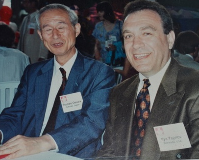
In the photo - with "the founding father of Cytochrome P450", prof. T. Omura (Congress in Toronto, Canada.1994).
It is a microsomal membrane-bound hemoprotein without known physiological functions at that time and was characterized by a unique 450-nm optical absorption peak of its carbon monoxide-bound form, which was originally reported as the spectrum of a novel “microsomal carbon monoxide-binding pigment” in 1958. Elucidation of its function as the monooxygenase in 1963 triggered a rapid expansion of research on this hemoprotein. Annual numbers of the published papers dealing with cytochrome P450, which were listed in Biological Abstracts and Citation Index, increased from 60 in 1970 to 500 in 1980, 900 in 1990, and 1500 in 1997. Cytochrome P450 is now regarded as the collective name of a large family of hemoproteins, “cytochrome P450 superfamily,” which seems to have diversified from a single ancestral protein to many forms during the course of biological evolution and is distributed widely among various forms of life from animals and plants to fungi and bacteria. Multicellular eukaryotic organisms including animals and plants have about 100 or more P450 genes in their genomes, and those many P450 genes are expressed tissue specifically and developmental stage specifically, indicating their diverse physiological functions. In mammals, various P450s participate in the biosynthesis and metabolism of sterols and steroid hormones and the metabolism of various lipid biofactors including eicosanoids, vitamin D3, and retinoids. Oxidative metabolism of foreign hydrophobic compounds as the first step of their excretion from the animal body is apparently another major function of cytochrome P450, which protects animals from noxious foreign compounds, man-created and natural.
 In the photo - major world players in the field of P450 studies (Ashford Castle, Ireland. 1978).
2.2. The 1970s: Studies on the monooxygenases as membrane-bound enzymes.
In the final year of medical school, Tsyrlov started working with a research group of Vyacheslav Lyakhovich (now has a RAS Academician rank in biochemistry) dealing with how lipid peroxidation of phospholipids in “dielectric” mitochondrial membrane affect oxidative phosphorylation activities. Later he applied the approach to “uncoupling” NADPH-dependent redox systems of the endoplasmic reticulum (microsomes), namely checked out structure-function relationships between integrity of microsomal phospholipids and catalytic parameters of membrane-bound monooxygenases (CYPs)[1]. The 50 human CYP molecular forms are all considered to be intrinsic membrane-bound proteins in the endoplasmic reticulum. Despite that in the 2000s it was showed that CYPs could be manipulated to yield crystals and structures, and that the modified protein was still catalytically active, evaluation of reactions catalyzed by “natural” CYPs embedded into a milieu of membrane phospholipids, is of importance, especially in cases when membranous phospholipid component would be modified due to intoxications or diseases’ development.
2.2.1.
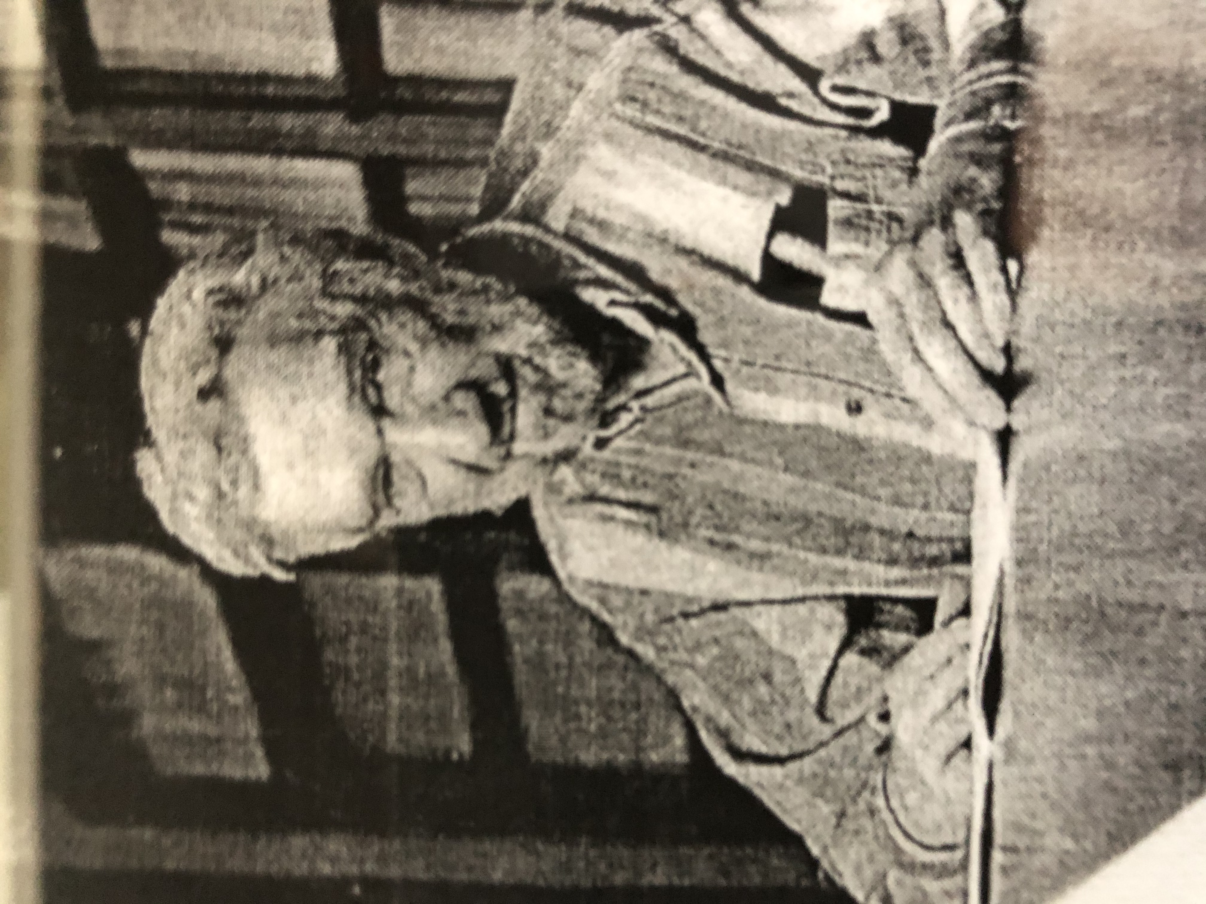
Tsyrlov's general research design in studies on experimental models or with epidemiology cohorts has always been to avoid using non-physiological (high) doses of any particular chemical agent, and to search for natural rout of administration for a chosen xenobiotic of interest. Thus administration to rodents in drinking water of industrial agent carbon tetrachloride (CCl4) at acute toxic doses resulted in activation of lipid-peroxidation reactions (LPR) in hepatic endoplasmic reticulum membranes followed by hepatocellular necrosis and fatal liver failure. Instead, while investigating chronic effects of CCl4 on hepatic microsomal monooxygenases, Tsyrlov used inhalation exposure of rats to this volatile agent at moderate doses[2,3]. A prolong inhalation by CCl4 of rats is regarded now as an accepted model of liver injury, similar to Laennec cirrhosis in man, which was morphologically determined by Vladimir Subbotin  (in the photo). Biochemically, it was found that a decreased p-hydroxylation of aniline during the entire period of liver cirrhosis development is determined by total amount of microsomal CO-binding hemoprotein. The rate-limiting step for N-demethylation of aminopyrine in the pre-cirrhotic period is the “substrate-binding” capacity of CYP2B1, while in microsomes from cirrhotic liver a “tight spot” in the metabolism of aminopyrine is the rate of an initial (fast) phase of NADPH-cytochrome P-450 reductase. Also, it was revealed for the first time that activation of LPR in liver microsomes from CCl4-cirrhotic and control rats caused the essentially different effects. In the controls, Tsyrlov with Vladimir Mishin with Mishin .pdf showed the peroxidative destruction of polyunsaturated arachidonic acid (C20:4) in microsomal phospholipids (a decrease of membrane hydrophobicity was measured by spectral, fluorometric and radiometric assays) and conversion of CYP2B1 to its inactive form. Alternatively to the controls, a pronounced resistance of microsomes from CCl4-cirrhotic rat liver to LPR activity was discovered. In all likelihood, microsomal phospholipids from cirrhotic liver contain mainly fatty acids with smaller amount of double bonds over that in control phospholipids. Such microsomal membrane structural readjustment was suggested a subcellular adaptation to long-term intoxication with pro-oxidant agent[4].
2.2.2.
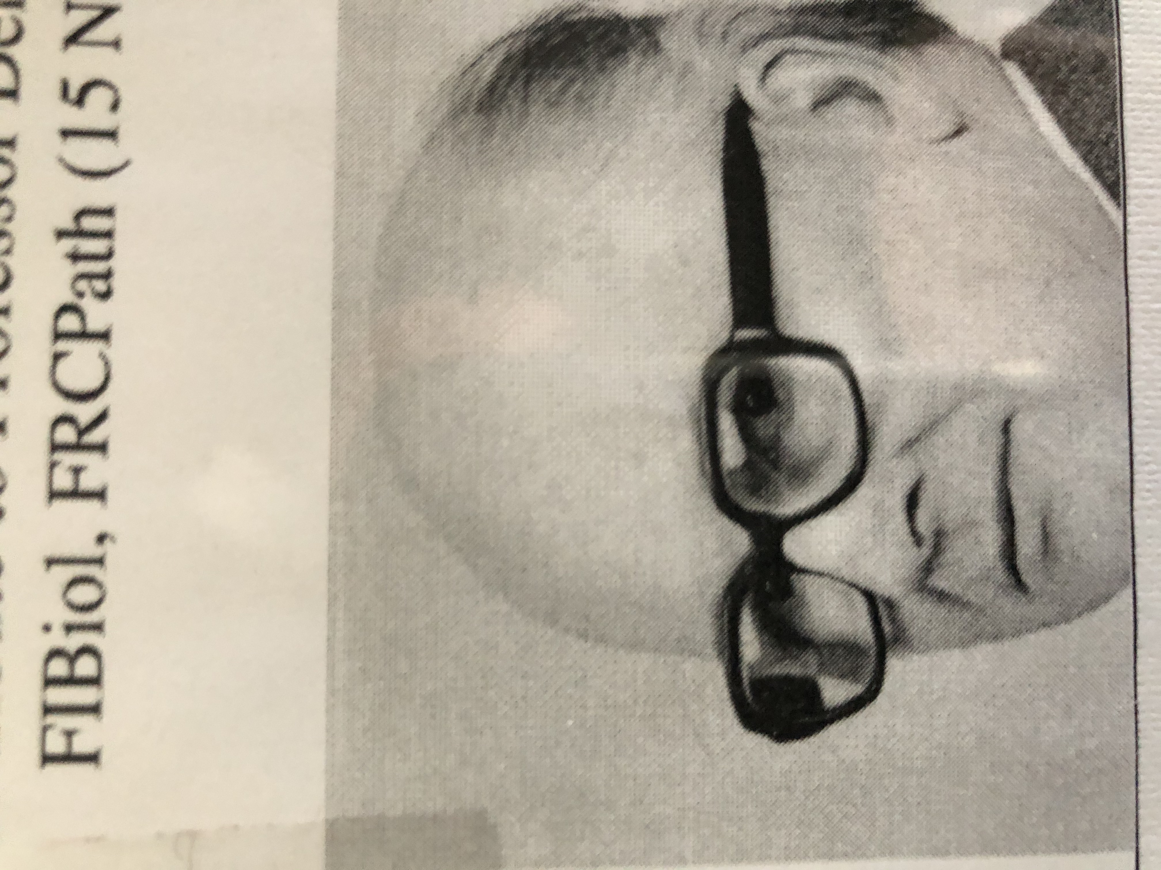
Earlier postulated essential requirements of phospholipids for electron transfer enzyme systems[5] include: decrease in the enzymic activity on removal of phospholipid, re-activation of the enzyme on addition of phospholipid, and association of enzyme with phospholipid. The Tsyrlov group addressed all the requirements, starting with the above models utilizing lipoperoxidative CCl4. Another approach of using the protonophore carbonyl cyanide m-chlorophenylhydrozone (CCCP) as a hydrophobic marker originated from studies of the future Nobel Prize winner (1978) Peter Mitchell (in the photo), who applied a lipophilic CCCP as uncoupling agent acting by transporting protons across the inner mitochondrial membrane[6].
.gif)
Dr. Mitchell supported the idea of using protonophores with “uncoupling” microsomal membrane, and thereafter communicated research results of Tsyrlov with Vyacheslav Lyakhovich and recently deceased Olga Gromova (in the photo)
 to The Biochemical Journal[7]. Factually, treatment of microsomes with 0.03—0,12 % deoxycholate or 10-90 μM CCCP reveals at least two different phospholipid-dependent hydrophobic zones assumed to exist in the microsomal membrane, both coupled with CYPs catalyzing N-demethylation of aminopyrine and p-hydroxylation of aniline. Increased concentrations of deoxycholate destroyed both zones. At the same time, as a less hydrophobic zone containing CYP’ haem group and binding sites for aniline, was not affected by the protonophore, a more hydrophobic zone containing the “substrate-binding protein” for aminopyrine and NADPH-cytochrome P-450 reductase, demonstrated selective specificity to the protonation action of CCCP. Our assumption of compartmentalized hydrophobic clusters in membrane phospholipids associated with different microsomal CYPs, was also proved together with Vladimir Mishin and Lev Wiener in experiments with an amide local anesthetic Cinchocain (brand names: Nupercaine and Sovcaine) [8], and with a highly lipophilic Cu(Lys)2 complex[9].
2.2.3.
Later, Tsyrlov with Nataly Zakharova-Polyakova N Polyakova.pdf quantitatively compared in vitro deoxycholate and cholate effects on microsomal monooxygenases with in vivo effects of accumulated dihydro and trihydro bile acids during cholestasis. The study has been highly recognized by a Member of Nobel Price Committee, Dr. Lars Ernster (in the photo, second from the left)  who communicated the paper to a reputable journal[10]. In fact, liver dysfunctions due to biliary duct obstruction are considered of special importance among non infectious liver diseases. To the point, hepatic microsomal CYPs were shown to play a double-edge role in cholestasis, as being an enzyme system responsible for oxidative metabolism of endogenous cholesterol and its hydroxylated derivatives, and at the same time CYP’ activities are a target for biliary acids due to their detergent action of membrane phospholipids. To test the concept of CYP’ phospholipid microenvironment, Tsyrlov with Nikolay Rivkind Kolya Rivkind Doc5.pdf studied hepatic monooxygenase systems reconstructed by means of self-assembly from rat [11] and murine[12] microsomes. For that, sodium cholate at critical micelle concentration was added and thereafter removed by dialysis thus allow a self-assembly of membrane proteins and bilayer-forming amphiphilic phospholipids to occur. Comparing to many widely known reconstituted monooxygenase systems, this one was characterized by the highest level of reactivation of catalytic activities due to preservation of CYP’ substrate-binding and active centers by glycerol at the stage of solubilization.
It is noteworthy that in the first-ever monograph on biochemistry of membrane-bound monoxygenases[13], Tsyrlov with Vyacheslav Lyakhovich With Lyakhovich.pdf presented massive literature data and own results on structural organization of microsomal phospholipids, and their regulatory role in every steps of CYP catalytic cycle during metabolism of xenobiotics (drugs, polycyclic hydrocarbons, etc) and endogenous substrates (steroids, fatty acids, etc.).
Special emphasis (quarter a century before David Lewis et al. came to the same conclusion[14]) was given on Corwin Hansch pioneer data on hydrophobic drugs with high octanol/water partition coefficients (log P) mainly distributed to hydrophobic areas such as phospholipid bilayers of cellular membrane[15]. Hansch approach was applied to CYP substrates thus assuming that substrate-binding affinity and catalytic activity of any individual CYP cannot be correctly evaluated without inclusion of substrate log P value.
REFERENCES
        [1] Omura, T., Sato, R. (April 1962) “A new cytochrome in liver microsomes”. The Journal of Biological Chemistry. 237:1375-1376.
        [2] Tsyrlov, I.B., Lyakhovich, V.V. (June 1972). “Damage effect of chronic intoxication by CCl4 on structural organization of liver microsomes and cytochromes (b)5 and P-450”. Biochemical Pharmacology. 21:2540-2542.
        [3] Tsyrlov, I.B., Lyakhovich, V.V. (May 1975). “ Rate-limiting steps in drug metabolism by microsomes from CCl4-cirrhotic rat liver”. Chemico-Biological Interactions 10:77-89.
        [4] Tsyrlov, I.B., Mishin, V.M., Lyakhovich, V.V. (November 1972). “Resistance of microsomes from CCl4-cirrhotic rat liver to lipoperoxidation activity”. Life Sciences 11(21):1045-1054. DOI: 10.1016/0024-3205(72)90206-8.
        [5] Fleischer, S., Brierley, G., Klouwen, H., Slautterback, D. B. (October 1962). “Studies of the electron transfer system. XLVII. The role of phospholipids in electron transfer”.  The Journal of Biological Chemistry. 237:3264-3272.
        [6] Mitchell, P. (February 1970). "Aspects of the chemiosmotic hypothesis". The Biochemical Journal. 116 (4): 5P–6P. DOI:10.1042/bj1160005p
        [7]  Tsyrlov, I.B., Gromova, O.A., Lyakhovich, V.V. (October 1976). “Mechanism of inhibition by carbonyl cyanide m-chlorophenylhydrozone and sodium deoxycholate of cytochrome P-450-catalyzed hepatic microsomal drug    metabolism”. The Biochemical Journal. 160(1):75-83. DOI:10.1042/bj1600075
        [8] Mishin, V., Tsyrlov, I., Gromova, O., Lyakhovich, V. (May 1974). “Phospholipids as cinchocaine action site during inhibition of lipoperoxidative reactions in rat liver microsomes”. Hoppe-Seyler’s Zeitschrift fur Physiologische Chemie 355:626-632.
        [9] Lyakhovich, V.V., Tsyrlov, I.B., Mishin, V.M., Weiner, L.M., Rumyantseva, G., Eremenko, S.I., Budker, V.G. (July 1979). “Activated forms of oxygen in metabolism of xenobiotics catalyzed by cytochrome P-450”. Acta Biologica et Medica Germanica 38:201-206. Berlin : Akademie-Verlag. (OCoLC)1027710611. ISSN: 0001-5318.
        [10] Tsyrlov, I.B., Zakharova-Polyakova, N.E., Gromova, O.A., Pospelova, L.N., Lyakhovich, V.V. (October 1978). “An in vivo – in vitro comparison of the effects of bile acids on the structural organization and functional activity of liver microsomal monooxygenases”. Experimental and Molecular Pathology. 20:131-143. DOI:10.1016/0014-4800(78)90033-3
        [11] Tsyrlov, I.B., Mishin, V.M., Gromova, O.A., Zakharova-Polyakova, N.E., Lyakhovich, V.V. (November 1977). “Preparation of a high-active microsomal monooxygenase system reconstituted by means of self-assembly”. Biochemical Pharmacology 26:2060-2163. DOI: 10.1016/0006-2952(77)90017-x PMID: 921816.
        [12] Lyakhovich, V.V., Tsyrlov, I.B., Gromova, O.A., Rivkind, N.B. ( (April 1978). “Activities of 3,4-benzpyrene hydroxylase systems reconstituted by means of self-assembly from control and 3-methylcholanthrene induced liver microsomes of “susceptible” and “unsusceptible” mice”. Biochemical and Biophysical Research Communications 81:1329-1335. DOI:10.1016/0006-291X(78)91281-0
        [13] Lyachovich, V.V., Tsyrlov, I.B. “Structural Aspects of Monooxygenases Biochemictry” (July 1978). Academic Press, Novosibirsk. Ed. Academician Rudolf Salganik. 242 pp. L:21003-743/686-78
        [14] Lewis, D.F., Jacobs, M.N., Dickins, M. “Compound lipophilicity for substrate binding to human P450s in drug metabolism”. (June 2004). Drug Discovery Today 9:530-537. PMID: 15183161
        [15] Hansch, C. (February 1972). “Quantitative relationships between lipophilic character and drug metabolism”, Drug Metabolism Reviews 1:1-14.
2.3. The 1980s: Discovery of "substrate-type" induction of CYP2B later applied to synthesis of new long-acting forms of medications

This discovery and applied results all originated from the Tsyrlov' Laboratory of Xenobiochemistry, Russian Academy of Sciences
2.3.1.
The induced de novo biosynthesis of microsomal monooxygenases catalyzing drug metabolism and bioactivation of environmental pollutants has important clinical consequences. It is reflected in almost exponential increase of publications on the subject, and nowadays induction of CYPs by phenobarbital reveals online about 40,000 results, by 3-methylcholanthrene (3-MC) – 118,000 results, and by 2,3,7,8-tetrachlorodibenzo-p-dioxin (TCDD) – 148,000 results (see next chapter). Working in a distant Siberia and seeking out own research niche in keen competition worldwide, Tsyrlov had asked and reciprocally received mentor's support from the most noted scientists in an area of cytochrome P450 induction, namely Herbert Remmer, Allan Conney, Harry Gelboin, Ronald Estabrook, Edward Bresnick, Dennis Parke, and Daniel Nebert.
Dr. Harry Gelboin, Head, Laboratory of Molecular Carcinogenesis, NCI/NIH. In 1990, Gelboin invited Tsyrlov to take position of Sr. Enzymologist in his lab. The Gelboin' parents had Belarusian roots, and his middle name was Victor.
Academician Ronald Estabrook. He was world renowned for his knowledge of enzymatic reactions related to toxicology and steroid hormone biosynthesis. In 1980, Ronald Estabrook endorsed Ilya Tsyrlov to join international team that started the ISSX.
Prof. Dennis Parke. An author of the founding book "Metabolism of Foreign Compounds", and the Editor-in-Chief of "Xenobiotica". In 1993, Parke promoted a speedy publication in his journal of innovative Tsyrlov & Pokrovsky' article on transactivation of HIV-1 with TCDD.
2.3.2.
Initially, Ilya Tsyrlov with Nataly Polyakova-Zakharova N Polyakova.pdf studied in vivo models of diminishing initial substrate binding of phenobarbital to the CYP, in order to establish the necessity of this step in the mechanism of monooxygenases induction. Data on a “consecutive induction”[16] and cholestasis[17] allow assuming that induction by phenobarbital of major form CYP2B6 is performed by the parent molecule of inducer rather than any products of its primary metabolism. Then, Tsyrlov with Alex Anshitz suggested the membrane factor involvement in the position-specific metabolism of structurally different substrates by another Family CYP2 Subfamily C Member 9, CYP2C9, which metabolizes phenobarbital.[18] In phenobarbital-induced rat liver microsomes, this CYP catalyzes the hydroxylation of aliphatic cyclohexane into 4-hydroxy-cyclohexane-1-one, and tetralin into tetralol. Microsomal monooxygenation of phenobarbital occurs on the side chain of its barbituric acid nucleus converting it into 4-hydroxyphenobarbital.[19] So, elevated monooxygenation of all substrates tested is strictly site specific and mimics the enzyme specificity for phenobarbital. As membraneous positioning of amphitropic CYP2C9 requires that substrate channel is facing the membrane hydrophobic core, it was concluded that inducer binding to CYP forms a specific enzyme-substrate complex. Membrane phospholipid/protein core “preserves” configuration of this complex and that determines site-specific metabolism for other CYP2C9 substrates. It was the first evidence for the membrane-bound enzyme of Daniel Koshland induced-fit hypothesis recognizing the conformational plasticity of enzymes, which mold themselves to the shape of the molecule.
2.3.3.
Proceeding further with the fact of extremely slow metabolism of phenobarbital-type inducers by the monooxygenase system,
 Tsyrlov with Konstantin Gerasimov (in the photo) ambitiously targeted conversion of aminopyrine, a typical substrate of CYP2B6, into inducer of this enzyme. That was achieved by site-directed modification of the position of aminopyrine molecule, which undergoes monooxygenation at the CYP2B6 active site. In the National/European Patent,[20] and a subsequent regular paper,[21] a novel pharmaceutical method was invented - the substitution of two methyl groups in the Aminopyrine -N(CH3)2 position by an isopropyl group, which resulted in a clear induction effect registered by spectral, kinetic methods, radiological tests, and immunochemistry assays. Also, later site-directed structure modifications with isopropyl groups were used while developing prolonged forms of Methadone, a synthetic opiate used to treat severe pain and narcotic drug addiction, and Artemisinin, an endoperoxide lactone now known as first-line natural antimalarial drug.[22]
REFERENCES
        [16] Tsyrlov, I.B., Zakharova, N.E., Gromova, O.A., Lyakhovich, V.V. (January 1976). “ Possible mechanism of induction of liver microsomal monooxygenase by phenobarbital”. Biochimica et Biophysica Acta 421:44-56. DOI: 10.1016/0304-4165(76)90168-9
        [17] Tsyrlov, I.B., Polyakova, N.E., Gromova, O.A. Rivkind, N.B., Lyakhochovih, V.V. (March 1979). “Cholestasis as an in vivo model for analysis of the induction of liver microsomal monooxygenases by sodium phenobarbital and 3-methylcholanthrene”. Biochemical Pharmacology 28:1473-1478. DOI: 10.1016/0006-2952(79)90460-X
        [18] Anshitz, A.G., Sokolovsky, V.D., Burilin, S.Yu., Lyakhovich, V.V., Tsyrlov, I.B. (January 1979). “Influence of inducer structure on catalytic activities of microsomal enzymes”. Kinetics and Catalysis (Science Press) 20(4):865-869. UDK: 577.15.02:577.158
        [19] Freudenthal, R. I., Carroll F. I. October 1973). “Metabolism of certain commonly used barbiturates”. Drug Metabolism Reviews 2:265–278. DOI:10.3109/03602537409030012
        [20] Tsyrlov, I.B., Gerasimov, K.G. (May 1986). “New Approach to Production of Prolong Acting Medicines Facilitating Hepatic Drug Detoxification System“. Patent SU/EP: 1457593 A1 4 G 01 N 33/48
        [21] Tsyrlov, I.B., Gerasimov, K.G. (June 1991). “Aminopyrine-N-demethylase I. Directed modification of substrates' structure as a way of production of inducer of the monooxygenase isoform P-450b”. European Journal of Drug Metabolism and Pharmacokinetics 16(3):207-212. DOI:10.1007/BF03189961
        [22] Tsyrlov, I.B., Woo, C.S.T., Martin, V.P., Shur, I.N. (April 2014). Modification of CYP2B6-mediated site of metabolism on Artemisinin molecule as a way to prolong its maximal antimalarial efficacy. International Journal of infectious diseases. Official publication of the International Society for Infectious Diseases 21(S1):174-177. DOI:10.1016/j.ijid.2014.03.784
2.3.4. Except for women from more vulnerable to malaria groups, a remedy hope for PCOS might be not Coartem® but rather long-acting formulations of oral Artemisinin
(Commentary article in Science, Jul. 12, 2024)

Ilya Tsyrlov (M.D., Ph.D., D.Sci) Chief Scientific Officer (former Research Professor at Icahn School of Medicine at Mount Sinai - NYC) Xenotox, Inc. (https://xenobiovir.com/) Irina Shur (M.D.) Internal Medicine Practitioner & a Medical Director, MEAC Montefiore Einstein Advanced Care (MEAC) of Montefiore Medical Center and Albert Einstein College of Medicine, Bronx, NY
The study by Liu et al. on ameliorating effects of artemisinins on polycystic ovarian syndrome (PCOS) via LONP-CYP11A1 interaction demonstrates potency of these medicines derived from Artemisia annua to be effective in improving the conditions of patients suffering from diseases other than multidrug resistant malaria (1). However, the authors did not consider two significant clinical issues. First, by choosing Coartem® with rodent models of PCOS and 19 PCOS patients, Liu et al. used Artemether in combination with Lumefantrine, a blood schizonticide agent of narrow specificity for erythrocytic stages of Plasmodium falciparum. This combination can cause side effects related to Coartem®, especially prolongation of the QT interval and arrhythmia (2). PCOS patients are often obese and insulin-resistant diabetics having advanced coronary artery disease, impaired left ventricular function and prolongation of the QT-interval, that is considered as risk factors for sudden cardiac death (3). Thus, using Coartem® could expose PCOS patients to additional adverse events. Second, the authors employed Coartem® regimen intended for acute treatment of paroxysmal stage of malaria (six doses over 3 days), notwithstanding that more people tended to fail tolerating treatment with Artemether‐Lumefantrine even when get tested with four‐dose regimen (4). So, an implicit question is, does Coartem® find a rationale to be a “remedy hope for PCOS”?
Artemisinins have demonstrated own therapeutic efficacy against human illnesses beyond malaria, including cancer, diabetes, atherosclerosis, viral infections, autoimmune diseases and neurodegenerative diseases. An ambiguous goal still is to extend an extremely short half-life (1-2 hours) of orally administered artemisinins that undergo the first-pass metabolism catalyzed by hepatic monooxygenases. In order to maintain therapeutically active artemisinins, some protection of their parent molecules against metabolism was provided in Artemether by means of a C H to C F substitution, or by designing of oil-soluble Artelinic acid synthesized from Artemisinin metabolite, dihydroartemisinin. Unfortunately, both Artemether and Artelinic acid demonstrated gastrointestinal and neurotoxicity. Another approach to Artemisinin short half-live in liver microsomes is due to its demethylation predominantly catalyzed by CYP2B6, although genetic variation contributes to a ~100 fold variation in human CYP2B6 protein level and activity. To the point, administration of the CYP2B6 inhibitors to human liver microsomes profoundly decreased Artemisinin disappearance rates (5). Previously, under similar circumstances of analgetic aminopyrine being N-demethylated by rat liver CYP2B6 into non-active products, we designed a substitution of two methyl groups in the aminopyrine -N(CH3)2 position with an isopropyl group thus creating steric hindrance at CYP active site resulted in preservation of aminopyrine molecule (6). Such an approach we applied recently to Isopropylartemisinin (IPAM) biosynthesized in Artemisia annua plant extracts from mevalonic acid via dimethylallyl and isopentenyl pyrophosphates, where dimethylallyl groups at pyrophosphate were substituted with isopropylallyl groups (7). IPAM retained an intact 1,2,4-trioxane ring incorporating an endoperoxide bridge, and was metabolized by human CYP2B6 12-13 times slower than parent Artemisinin. The amounts of hydroxylated and glucuronide-conjugated derivatives of covalent heme adducts were equally high in urine of Plasmodium infected mice treated with Artemisinin or IPAM compared to Artemisinin metabolites lacking trioxane ring endoperoxide bridge. As artemisinins were also reported to have antiviral activity, Artemisinin and IPAM were tested as inhibitors in the human monocytic line infected with cytomegalovirus or its GCV-resistant strain. It was found that 0.5 mM IPAM provided >80% inhibition of GCV-resistant strain, while 5.0 mM Artemisinin caused ~40% plague reduction.
The article on ameliorating effects of Coartem® (1), and a response to the article (8) both referred to women/girls with PCOS from East/Southeast Asia regions with malaria transmission. Since PCOS is a chronic endocrine disorder widespread all over the world, it is recommended that in malaria-free countries the therapy for PCOS would be provided not with a fixed-dose combination of Artemether and Lumefantrine but rather with durable and tolerable Artemisinin derivatives designed to undergo slow presystemic metabolism.
References
- J. Liu, S.Y. Jiang et al., Artemisinins ameliorate polycystic ovarian syndrome by mediating LONP1-CYP11A1 interaction. Science 384, eadk5382 (2024). doi: 10.1126/science.adk5382
- Artemether And Lumefantrine (Oral Route) Side Effects https://www.mayoclinic.org/drugs-supplements/artemether-and-lumefantrine-oral-route/side-effects/drg-20072694. Last updated: June 01, 2024.
- Scicchitano et al., Cardiovascular risk in women with PCOS. Int. J. Endocrinol. Metab. 10(4):611–618 (2012). doi: 10.5812/ijem.4020
- Omari et al., Artemether‐lumefantrine (four‐dose regimen) for treating uncomplicated falciparum malaria. Cochrane Database Syst. Rev. 2:CD005965 (2006). doi:10.1002/14651858.CD005965
- S. Svensson and M. Ashton. Identification of the human cytochrome P450 enzymes involved in the in vitro metabolism of artemisinin. Br. J. Clin. Pharmacol. 48(4):528-35 (1999). doi:10.1046/j.1365-2125.1999.00044.x.
- I.B. Tsyrlov and K.E. Gerasimov. Aminopyrine-N-demethylase I. Directed modification of substrates’ structure as a way of production of inducer of the monooxygenase isoform P-450b. Eur. J. Drug Metab. Pharmacokinet.16, 207–212. (1991). doi.org/10.1007/BF03189961
- B. Tsyrlov, K.E. Gerasimov and I.N. Shur. Site-specific isopropylation of parent molecule as a way to develop artemisinin long-acting antimalarial and anticytomegalovirus formulation. J. Biol. Chem. 300, S21:1260. (2024). doi.org/10.1016/j.jbc.2024.105825
- de Zegher and L. Ibanez. Artemisinins for polycystic ovary syndrome: more than one mediating mechanism. Science 384, eLetter (2024)
JUL. 1, 2024. Re. Artemisinins ameliorate polycystic ovarian syndrome by mediating LONP1-CYP11A1 interaction. By Yang Liu et al., Science, 384, 14 JUN 2024 (eLetter to _Science _Editor)
ILYA TSYRLOV, CSO, Xenotox, Inc., 26 Shepherds Drive, Scarsdale, NY 10583, USA ([email protected]) IRINA SHUR, M.D. Physician (Primary Care), Montefiore Einstein Advanced Care, 555 Taxter Road, Elmsford, NY 10523, USA ([email protected])
Dear Editor,
The study by Liu et al. on ameliorating effects of artemisinins on polycystic ovarian syndrome (PCOS) via LONP-CYP11A1 interaction demonstrates potency of these medicines derived from Artemisia annua to be effective in improving the conditions of patients suffering from diseases other than multidrug resistant malaria. However, the authors did not consider two significant clinical issues. First, by choosing Coartem® with rodent models of PCOS and 19 PCOS patients, Liu et al. used Artemether in combination with Lumefantrine, a blood schizonticide agent of narrow specificity for erythrocytic stages of Plasmodium falciparum. This combination can cause side effects related to Coartem®, especially prolongation of the QT interval and arrhythmia (1). PCOS patients are often obese and insulin-resistant diabetics having advanced coronary artery disease, impaired left ventricular function and prolongation of the QT-interval, that is considered as risk factors for sudden cardiac death (2). Thus, using Coartem® could expose PCOS patients to additional adverse events. Second, the authors employed Coartem® regimen intended for acute treatment of paroxysmal stage of malaria (six doses over 3 days), notwithstanding that more people tended to fail tolerating treatment with Artemether‐Lumefantrine even when get tested with four‐dose regimen (3). So, an implicit question is, does Coartem® find a rationale to be a “remedy hope for PCOS”? Artemisinins have demonstrated own therapeutic efficacy against human illnesses beyond malaria, including cancer, diabetes, atherosclerosis, viral infections, autoimmune diseases and neurodegenerative diseases. An ambiguous goal still is to extend an extremely short half-life (1-2 hours) of orally administered artemisinins that undergo the first-pass metabolism catalyzed by hepatic monooxygenases. In order to maintain therapeutically active artemisinins, some protection of their parent molecules against metabolism was provided in Artemether by means of a C-H to C-F substitution, or by designing of oil-soluble Artelinic acid synthesized from Artemisinin metabolite, dihydroartemisinin. Unfortunately, both Artemether and Artelinic acid demonstrated gastrointestinal and neurotoxicity. Another approach to Artemisinin short half-live in liver microsomes is due to its demethylation predominantly catalyzed by CYP2B6, although genetic variation contributes to a ~100 fold variation in human CYP2B6 protein level and activity. To the point, administration of the CYP2B6 inhibitors to human liver microsomes profoundly decreased Artemisinin disappearance rates (4). Previously, under similar circumstances of analgetic aminopyrine being N-demethylated by rat liver CYP2B6 into non-active products, we designed a substitution of two methyl groups in the aminopyrine -N(CH3)2 position with an isopropyl group thus creating steric hindrance at CYP active site resulted in preservation of aminopyrine molecule (5a). Such an approach we applied recently to Isopropylartemisinin (IPAM) biosynthesized in Artemisia annua plant extracts from mevalonic acid via dimethylallyl and isopentenyl pyrophosphates, where dimethylallyl groups at pyrophosphate were substituted with isopropylallyl groups (5b). IPAM retained an intact 1,2,4-trioxane ring incorporating an endoperoxide bridge, and was metabolized by human CYP2B6 12-13 times slower than parent Artemisinin. The amounts of hydroxylated and glucuronide-conjugated derivatives of covalent heme adducts were equally high in urine of Plasmodium infected mice treated with Artemisinin or IPAM compared to Artemisinin metabolites lacking trioxane ring endoperoxide bridge. As artemisinins were also reported to have antiviral activity, Artemisinin and IPAM were tested as inhibitors in the human monocytic line infected with cytomegalovirus or its GCV-resistant strain. It was found that 0.5 mM IPAM provided >80% inhibition of GCV-resistant strain, while 5.0 mM Artemisinin caused ~40% plague reduction. Referring to the study by Liu et al., since PCOS is a chronic endocrine disorder that has no cure, it is recommended that the short ameliorating effect of Coartem® on PCOS be improve by using more durable and tolerable Artemisinin derivatives with slow first-pass metabolism mediated by CYP monooxygenases.
References
1) Artemether And Lumefantrine (Oral Route) Side Effects https://www.mayoclinic.org/drugs-supplements/artemether-and-lumefantrine-oral-route/side-effects/drg-20072694. Last updated: June 01, 2024.
2) Scicchitano et al., Cardiovascular risk in women with PCOS. Int. J. Endocrinol. Metab. 10(4):611–618 (2012). doi: 10.5812/ijem.4020
3) Omari et al., Artemether‐lumefantrine (four‐dose regimen) for treating uncomplicated falciparum malaria. Cochrane Database Syst. Rev. 2:CD005965 (2006). doi:10.1002/14651858.CD005965
4) S. Svensson and M. Ashton. Identification of the human cytochrome P450 enzymes involved in the in vitro metabolism of artemisinin. Br. J. Clin. Pharmacol. 48(4):528-35 (1999). doi:10.1046/j.1365-2125.1999.00044.x.
5a) I. B. Tsyrlov and K.E. Gerasimov. Aminopyrine-N-demethylase I. Directed modification of substrates’ structure as a way of production of inducer of the monooxygenase isoform P-450b. Eur. J. Drug Metab. Pharmacokinet.16, 207–212. (1991). doi.org/10.1007/BF03189961;
5b) I.B. Tsyrlov et al., Site-specific isopropylation of parent molecule as a way to develop artemisinin long-acting antimalarial and anticytomegalovirus formulation. J. Biol. Chem., 300, S21:1260. (2024). doi.org/10.1016/j.jbc.2024.105825
2.4. The 1990s: Human studies using the recombinant CYPs as preferred tests for major medical conditions
2.4.1.
As an enzymologist with expertise in comparative analyses of human and mouse CYP1As, including development of the "mutual depletion inhibition system",[39] Tsyrlov got an offer from Harry Gelboin to investigate the enzyme kinetics of the recombinant human and murine CYP1As expressed through the vaccinia virus system in human hepatoma HepG2 cells developed by outstanding Frank Gonzalez' team.
Dr. Frank J. Gonzalez is currently a Head of Laboratory of Metabolism, Center for Cancer Research, NCI/NIH. He is the world leading scientist in the field of CYP genes regulation.
Tsyrlov led the NCI Subproject.[40] In 1993, the first-ever published enzyme-kinetics comparative analysis was applied to hepatic microsomal, purified, and the recombinant Cyp1a1 and Cyp1a2 from mice.[41] Usage the recombinant CYPs as the markers of metabolic drug-drug interaction is now a typical feature for many standard medical (PDR), toxicology, and oncology handbooks. Particularly, Tsyrlov with Harry Gelboin determined the [[Km]] and [[Kcat]] values thus revealing substrate specificity and catalytic activity for mouse Cyp1a1 and Cyp1a2 expressed through the vaccinia virus system in human hepatoma HepG2 cells.
The results demonstrated an advantage of the recombinant CYP1As over their counterparts in microsomal and purified reconstructed systems. Activities of the recombinant mouse Cyp1a1, Cyp1a2, and human CYP1A2 and CYP2B6 amplified in HepG2 and TK- cells if added with human cDNA-expressed P450-oxidoreductase, at optimal ratio the oxidoreductase to CYP of 0.75, and inhibitory effects of isozyme-specific monoclonal antibodies were stronger after co-expression. With embryoblast TK- cells expressing low-active recombinant CYPs, the co-expression led to a significant increase of Kcat values without any change of Km. Partial inactivation of cDNA-expressed human CYP2E1 in the host cell was protected by the addition of imidazole into the culture medium and subsequent washing out inhibitor after cells harvesting.
2.4.2.
The study of Tsyrlov with Victor Michailenko (in the photo)  concerned the mechanisms of effects of bioflavonoids on the recombinant murine and human CYP1A2s,[42] and by far preceded multiple other publications on the subject. Bioflavonoids, natural substances with variable structures, are found in fruits, vegetables, grains, bark, roots, stems, flowers, tea, and wine. The inhibitory effects on CYP1As of several bioflavonoids were studied in relation to the position of hydroxyl groups on the rings. A comparison between the mouse Cyp1a2 and human CYP1A2s revealed species-specific differences, although hydroxylated flavonoids inhibited the AHH activity of both CYP1As due to suppression of the NADPH-cytochrome P450 oxidoreductase. Structure-activity relationships showed the importance of hydroxyl groups at 5 and 7-positions on the ring A of flavone nucleus exemplified by the strongest inhibitory potency of Chrisin. The presence of a hydroxyl group at the 4’-position on the ring B decreased the inhibitory effect of a bioflavonoid. The ortho-orientation of a hydroxyl group on the ring C was of importance, as Quercetin inhibits cDNA-expressed and hepatic CYP1s more effectively than Morin. Bioflavonoids that selectively inhibit the CYP1As may be utilized as chemoprotective remedies to reduce the risk of certain cancers. The latter approach was also chosen by Tsyrlov with Valerian Kagan (in the photo)
  using purified recombinant human NADPH-cytochrome P450 oxydoreductase in the study of metabolism-based toxicity of phenoxyl radical of a phenolic antitumor drug Etoposide.[43]
2.4.3.
An interest in studying the CYP1A2 evolved because this unique CYP, by Fred Guengerich, “oxidizes both endogenous and xenobiotic substrates, like centaurs in Greek mythology, who had the upper body of a human fused to the body of a horse, as it … operates in two worlds at once doing the same chemistry in both worlds”.
Prof. Fred P. Guengerich is currently a leading scientist in the field of basic and applied studies on the CYP-substrate relationships
As regards medically related studies, Tsyrlov' revealing information on the microsomal and recombinant mouse Cyp1a2 is among the principal publications in UniProtKB - P00186 (CP1A2_MOUSE) global database (https://www.uniprot.org/uniprot/P00186). Tsyrlov with Peter Sinclair (in the photo)

evaluated what human CYP predominantly catalyzes uroporphyrinogen oxidation.[44] Porphyria is a group of diseases in which porphyrins build up, negatively affecting the skin or nervous system. Complications of acute porphyria attacks include paralysis, low blood sodium levels, seizures, and such attacks may be triggered by alcohol and smoking. In previous studies on the oxidation of uroporphyrinogen to uroporphyrin in human liver microsomes, CYP1A2 was found relatively weak compared to mouse liver microsomal Cyp1a2, and the CYP2E1 and CYP3A4 were not studied at all. So, uroporphyrinogen oxidation along with isozyme-specific reactions for the recombinant CYP1A2, CYP2E1, and CYP3A4 expressed in HepG2, TK- cells and Escherichia coli were tested. It was concluded that only a small proportion of uroporphyrin formation in human liver microsomes belongs to CYP1A2, although human CYP1A2 can support uroporphyrin accumulation in hepatoma cells. In another study, Tsyrlov with Katja Salmela (in the photo)

investigated the involvement of human CYP1A2 and CYP3A4 as possible constituents of the microsomal ethanol oxidizing system (MEOS).[45] MEOS is an alternative to alcohol dehydrogenase pathway of ethanol oxidation to acetaldehyde, and in MEOS the dominant role had been attributed to the CYP2E1.[46] MEOS is involved in many kinds of diseases, such as alcoholic liver disease and diabetes. Although ethanol-induced CYP2E1 plays an important role in the xenobiotic metabolism and chemical toxicity, few animal models are used to predict CYP2E1 properties in physiology, pathology, as well as activation.
 Prof. Charles Lieber was an inventor of the term MEOS, and a world-leading scientist in the field of alcohol liver disease.
Regarding the CYP1A2, its noteworthy this CYP can be induced by cigarette smoking, and the CYP3A4 is a catalyst of many monooxygenase reactions involved in drug metabolism and synthesis of cholesterol, steroids, and other lipids. To establish a novel model for investigating the functions of all three human CYPs in ethanol oxidation, enzyme kinetics, and inhibitory analyses of human hepatoma-derived HepG2 cells overexpressing the recombinant human CYP2E1, CYP1A2 or CYP3A4 were utilized. All three recombinant CYPs metabolize ethanol, and the most catalytically active was CYP1A2, followed by CYP2E1 and CYP3A4. However, in human liver microsomes, due to the lowest immunochemically determined content of CYP2E1, its ethanol-oxidizing catalytic activity was 60% and 230% higher than that of CYP1A2 and CYP3A4, respectively. In terms of total activity (nmol acetaldehyde/min x nmol CYP), the scale was: CYP3A4 (the most abundant hepatic CYP) followed by CYP2E1 and CYP1A2. In normal human liver, a dominance of total CYP3A4 ethanol-oxidizing activity is even stronger, because CYP3A4 content is determined over 40% of all liver centrilobular CYPs, whereas the contents of CYP1A2 and CYP2E1 constituted 18% and 9%, respectively. As heavy drinking of alcohol is commonly associated with heavy smoking, their synergistic effects have been often described in clinics. So, the possibility of an additive effect of simultaneous CYP2E1 and CYP1A2 inductions was tested in liver microsomes of rats treated with ethanol and/or 3-MC.[47] Rats were fed the Lieber-Carli liquid ethanol diet for 3 weeks and treated by 3-MC for 5 days. Enzyme kinetics analysis revealed that combined treatment of rats with ethanol and 3-MC significantly diminishes the induction of CYP2E1 by ethanol, and despite its low catalytic activity, 3-MC-induced was shown significantly contributing to microsomal ethanol oxidation to acetaldehyde. Therefore, by all means, the CYP1A2 and CYP3A4 might be considered active components of MEOS. As concerns CYP1A2 gene activation by xenobiotics, it is characterized by unusual patterns different from activation of the CYP1A1 gene. Such a case was exemplified by acenaphthylene, a tricyclic PAH formed as a by-product of incomplete combustion of fossil fuels. Using immunoinhibitory, spectral, enzyme kinetics methods, nuclear run-on assay and the AhR density gradient analysis, Tsyrlov with Steven Safe revealed that acenaphthylene and related tricyclic PAHs could induce Cyp1a2 gene product in mice of genotype Ahb/Ahd without binding to the AhR.[48]
REFERENCES
        [39]  Tsyrlov, I.B., Lyakhovich, V.V. (September 1978). "Genetic variations in cytochrome P-450 catalytic center activities in relation to subsequent conjugation reactions". In: Conjugation Reactions in Drug Biotransformation, Ed. A. Aitio, Elsevier/North-Holland Biomedical Press, Amsterdam-New York, Oxford. pp.135-145. ISBN:0-444-80090-5
        [40] Tsyrlov, I.B., Gonzalez, F.J., Korzekwa, K.R., Mikhailenko, V.M., Gelboin, H.V. (February 1993). “Isozyme-specific inhibition of cDNA-expressed and liver microsomal P450s”. Summary of Research 1990-1993. Chemical and Physical Carcinogenesis Program. Division of Cancer Etiology. NCI/NIH. 66-76. ref.https://dceg.cancer.gov/
        [41] Tsyrlov, I.B., Goldfarb, I.S., Gelboin, H.V. (December 1993). “Enzyme-kinetic and immunochemical characteristics of mouse cDNA-expressed, microsomal, and purified CYP1A1 and CYP1A2”. Archives of Biochemistry and Biophysics 307:259-266. DOI:10.1006/abbi.1993.1588
        [42] Tsyrlov, I.B., Mikhailenko, V.M., Gelboin, H.V. (March 1994). “Isozyme- and species-specific susceptibility of cDNA-expressed CYP1A P-450s to different flavonoids”. Biochimica et Biophysica Acta 1205:325-335. DOI: 10.1016/0167-4838(94)90252-6
        [43] Goldman, R., Tsyrlov, I.B., Grogan, J., Kagan, V.E. (March 1997). “Reaction of phenoxyl radicals with NADPH-cytochrome P-450 oxidoreductase and NADPH: reduction of the radicals and inhibition of the enzyme”. Biochemistry 36:3186-3192. DOI: 10.1021/bi9621728
        [44] Sinclair, P.R., Gorman, N., Tsyrlov, I.B., Fuhr, U., Walton, H.S., Sinclair, J.F (October 1998). “Uroporphyrinogen oxidation catalyzed by human cytochromes P450”. Drug Metabolism and Disposition 26:1019-1025. Print ISSN: 0090-9556. PubMed: 9763408A
        [45] Salmela, K.S., Kessova, I.G, Tsyrlov, I.B., Lieber, C.S. (December 1998). “Respective roles of human cytochrome P4502E1, 1A2 and 3A4 in the hepatic microsomal ethanol oxidizing system”. Alcoholism: Clinical and Experimental Research 22:2125-2132. DOI: 10.1111/j.1530-0277.1998.tb05926
        [46] Lieber, C.S. (October 2004). “The Discovery of the Microsomal Ethanol Oxidizing System and Its Physiologic and Pathologic Role”. Drug Metabolism Reviews 36:511-529. DOI: 10.1081/dmr-200033441
        [47] Tsyrlov, I.B., Salmela, K.S., Lieber, C.S. (December 1996). “Inhibitory effect of 3-methylcholanthrene on hepatic microsomal ethanol inducible cytochrome CYP2E1 in rats”. Mount Sinai Journal of Medicine. A Journal of Translational and Personalized Medicine 64(1):30-31. ISSN:0027-2507
        [48] Chaloupka, K., Santostefano, M., Goldfarb, I.S., Liu, G., Myers, M.J., Tsyrlov, I.B., Gelboin, H.V., Krishnan, V., Safe, S. (December 1994). “Aryl hydrocarbon (Ah) receptor-independent induction of Cyp1a2 gene expression by acenaphthylene and related compounds in B6C3F1 mice”. Carcinogenesis 15:2835-2840. DOI:10.1093/carcin/15.12.2835
At the end of the day, we all are people devoted our careers (and our cars🤠) to the field of P450
(In the photo - meeting in New York with a former lab colleague, prof. Lyudmila Gulyaeva)